🦷 DR FARHAN EXPLAINS — CLINICAL SERIES
Biopsy in the Oral Cavity — Stepwise Protocol
Lesion Selection, Technique Algorithms & Safety Decision Guide
FRONT MATTER
Series Branding
Dr Farhan Explains — Clinical Series
Algorithm-Driven Clinical Guides for Oral Medicine & Minor Oral Surgery
Professional Foreword
Biopsy is the definitive diagnostic procedure for suspicious oral lesions — yet clinical failures often arise from wrong site selection, wrong biopsy type, poor margin choice, or delayed referral. The technical act of cutting tissue is simple; the decision architecture behind biopsy is not.
Correct biopsy practice requires structured decisions on:
Whether to biopsy
When to biopsy
Where to biopsy
Which biopsy type to use
When to refer instead
How to document and transport tissue
This guide converts oral biopsy into a clinical decision and execution algorithm system suitable for dentists, orthodontists, oral physicians, and postgraduate trainees.
This is a protocol and safety manual — not a surgical atlas.
How To Use This Guide
Apply in sequence:
1️⃣ Lesion risk screening
2️⃣ Biopsy indication filter
3️⃣ Contraindication check
4️⃣ Biopsy type selection
5️⃣ Site selection algorithm
6️⃣ Stepwise procedural protocol
7️⃣ Specimen handling rules
8️⃣ Report & follow-up pathway
Each section includes:
Flowchart logic
Threshold triggers
Safety rules
Medicolegal checkpoints
Examiner viva notes
Pitfalls
Icon Legend
🔴 Red Flag — urgent biopsy/referral
🦷 Chairside Tip — technical execution detail
⚠️ Medicolegal — documentation trigger
📘 Examiner Note — viva anchor
❌ Pitfall — common biopsy error
SECTION 1 — Biopsy Indication Master Algorithm
Decision Flow — Should You Biopsy?
Oral lesion detected
↓
Persists > 2 weeks?
↓
YES
↓
Unknown diagnosis?
OR
OPMD suspicion?
OR
Induration/ulcer/red lesion?
↓
YES → Biopsy indicated
Automatic Biopsy Indications
🔴 Non-healing ulcer >2 weeks
🔴 Red or red-white lesion
🔴 Indurated lesion
🔴 Suspicious leukoplakia
🔴 Erosive OLP (atypical)
🔴 Pigmented lesion (new/irregular)
🔴 Unexplained swelling
📘 Examiner Note: Persistence + uncertainty = biopsy
SECTION 2 — Do NOT Biopsy First — Referral First Situations
Immediate Specialist Referral Instead
Highly vascular lesion
Suspected malignancy large
Deep floor of mouth lesion
Base of tongue lesion
Bleeding disorder patient
🔴 Red Flag: Large suspected carcinoma → refer, not chairside biopsy
SECTION 3 — Biopsy Type Selection Algorithm
Biopsy Type Decision Tree
Small lesion (<1 cm)?
→ Excisional biopsy
↓
Large lesion?
→ Incisional biopsy
↓
Diffuse mucosal disease?
→ Incisional representative biopsy
↓
Fluid lesion?
→ Aspiration first
Biopsy Type Matrix
| Type |
Use |
| Excisional |
Small benign lesion |
| Incisional |
Large/suspicious lesion |
| Punch |
Flat mucosal lesion |
| Aspiration |
Fluctuant/swelling |
SECTION 4 — Incisional vs Excisional Rules
Excisional Criteria
<1 cm
Clinically benign
Accessible
Low malignancy suspicion
Incisional Criteria
1 cm
Suspicious features
Mixed color lesion
Induration present
❌ Pitfall: Excisional biopsy of suspected carcinoma
SECTION 5 — Where to Biopsy — Site Selection Algorithm
Site Selection Flow
Heterogeneous lesion?
↓
Sample most abnormal area
↓
Include red/indurated zone
↓
Avoid necrotic center
↓
Include margin of normal tissue
Correct Target Areas
| Lesion Type |
Biopsy Site |
| Red-white |
Red component |
| Ulcer |
Edge, not center |
| Large plaque |
Thickest change |
| Mixed |
Most dysplastic-looking |
🦷 Chairside Tip: Edge = highest diagnostic yield
❌ Pitfall: Sampling only keratinized white surface
SECTION 6 — Oral Biopsy Instruments & Setup
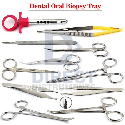
Standard Setup
Scalpel blade (15 preferred)
Tissue forceps (non-crushing)
Suture kit
Formalin container
Gauze & suction
Local anesthetic (without lesion injection)
SECTION 7 — Anesthesia Protocol
Injection Rule
Inject around lesion — not into lesion
Reason:
Avoid artifact
Avoid tumor cell distortion
Preserve histology
🦷 Chairside Tip: Field block preferred
❌ Pitfall: Infiltrating directly into lesion
SECTION 8 — Stepwise Biopsy Procedure (Incisional)
Procedural Algorithm
Consent obtained
↓
Photo & measurement
↓
Mark biopsy site
↓
Field anesthesia
↓
Elliptical incision
↓
Depth into submucosa
↓
Lift with forceps edge
↓
Cut base cleanly
↓
Place in formalin immediately
↓
Suture
SECTION 9 — Specimen Size Rules
Minimum Size
| Dimension |
Target |
| Length |
≥5 mm |
| Depth |
Include epithelium + connective tissue |
📘 Examiner Note: Too small = nondiagnostic
SECTION 10 — Handling & Fixation Protocol
Fixation Rules
Specimen → immediately into 10% formalin
Volume = 10× tissue volume
Label before procedure
Label Must Include
Patient ID
Site
Date
Clinician
Clinical description
⚠️ Medicolegal: Mislabeling = critical error
SECTION 11 — Special Site Modifications
Tongue / Floor of Mouth
Higher bleeding risk
Deeper vessels
Consider referral if large
Gingiva
Include epithelium + connective tissue
Avoid periosteal stripping if not needed
SECTION 12 — Punch Biopsy Algorithm
Use Punch When
Flat mucosal lesion
Immune disease suspected
OLP / vesiculobullous disease
Rotate punch
↓
Lift core
↓
Cut base
❌ Pitfall: Punch on indurated carcinoma (too shallow)
SECTION 13 — Vesiculobullous Disease Biopsy Rule
Dual Biopsy Protocol
One biopsy — lesion edge (H&E)
One biopsy — perilesional normal (DIF)
📘 Examiner Note: DIF requires special medium (not formalin)
SECTION 14 — Hemostasis & Closure
Hemostasis Methods
Pressure
Sutures
Local hemostatic agents
Avoid:
Excess cautery at margin (histology damage)
SECTION 15 — Post-Op Care Protocol
Patient Instructions
No trauma to site
Soft diet
Avoid rinsing 24 hrs
Return if bleeding
SECTION 16 — Pathology Request Form — Mandatory Fields
Must Include
☑ Duration
☑ Site
☑ Size
☑ Risk factors
☑ Clinical impression
☑ Photo reference
⚠️ Medicolegal: “Clinical correlation required” depends on your notes
SECTION 17 — Result Follow-Up Algorithm
Report received
↓
Benign → treat/monitor
Dysplasia → specialist referral
Malignancy → urgent oncology referral
Inconclusive → repeat biopsy
❌ Pitfall: Not reviewing pathology report personally
SECTION 18 — Examiner Viva High-Yield Points
📘 Biopsy = gold standard diagnosis
📘 Edge of ulcer — not center
📘 Red area preferred
📘 Inject around, not into lesion
📘 Incisional for suspicious lesions
📘 Formalin immediately
📘 DIF needs special transport
SECTION 19 — Clinical Pitfalls
❌ Biopsying necrotic center
❌ Too superficial sample
❌ Crushing tissue with forceps
❌ Thermal artifact from cautery
❌ No photo record
❌ No pathology follow-up
APPENDICES — POSTER & WORKSHEET PACK
Poster Pack (A3/A4 Ready)
1️⃣ Oral Biopsy Decision Flowchart
2️⃣ Incisional vs Excisional Tree
3️⃣ Site Selection Guide
4️⃣ Biopsy Step Protocol Poster
5️⃣ Specimen Handling Rules
6️⃣ DIF Dual Biopsy Poster
7️⃣ Contraindication Checklist
8️⃣ Result Follow-Up Algorithm
Chairside Worksheets
Biopsy Indication Checklist
Site Selection Map Sheet
Specimen Label Template
Pathology Request Form Guide
Follow-Up Tracking Log
REFERENCES — VANCOUVER STYLE
Neville BW, Damm DD, Allen CM. Oral and Maxillofacial Pathology.
Regezi JA, Sciubba JJ, Jordan RCK. Oral Pathology.
Hupp JR, Ellis E, Tucker MR. Contemporary Oral & Maxillofacial Surgery.
WHO Head & Neck Tumour Classification.
Major oral medicine biopsy guidelines.
 Emergency Dental Care